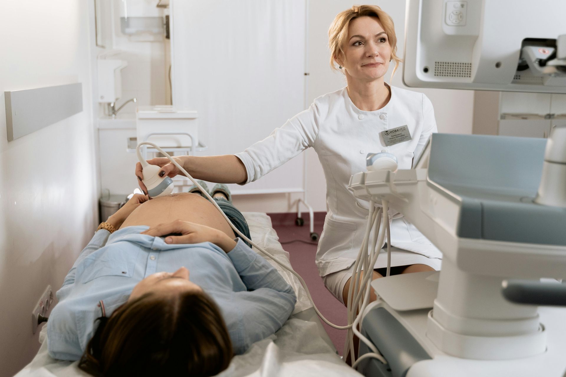
Routine Ultrasound Often Misses Endometriosis
Learn More about Ultrasound Recommendations to Improve Detection of Superficial and Deep endometriosis, and Endometrial Pelvic Scar Tissue Adhesions

Endometriosis has been associated with infertility and subfertility, affecting up to 50% of patients with fertility issues. [1]
In recent years, increased understanding of the disease has suggested that endometriosis is associated with infertility and subfertility affecting up to as many as 90% of patients with fertility issues. [2]
In the U.S., there is an average over seven-year delay between the onset of symptoms and a diagnosis of endometriosis. [3, 4]
In the U.S., at the time of this writing, endometriosis diagnosis can only be confirmed by a surgical procedure--typically a minimally invasive laparoscopy; however, doctors often recommend doing medical imaging first.
Routine Pelvic Ultrasound for Endometriosis
The first line of medical imaging used to identify endometriosis is routine pelvic ultrasound. However, it often misses the most severe form of endometriosis--that is, hidden endometriosis growing inside pelvic organs instead of outside them. This most severe form of endometriosis can extend to any depth beneath the peritoneal surface [5], and is known as deep endometriosis (DE) and deep infiltrating endometriosis (DIE).
Another common sign of endometriosis commonly missed on routine pelvic ultrasound is adhesions. Adhesions, often simply referred to as scar tissue, cause pelvic organs to be tethered or fused together. Tethering and fusion of organs often impairs their proper movement and function. Common organs affected by endometrial fusion and tethering include the cervix, uterus, fallopian tubes, ovaries, bladder, colon, and appendix . . . along with muscles and nerves.
Other than the visualization of an endometrioma, sonologists who perform routine pelvic transvaginal ultrasound for pelvic pain are often not identifying endometriosis. [6]
In one retrospective study published in 2020, simple routine ultrasound was performed on 150 women with chronic pelvic pain, and nothing abnormal was detected. However, follow-up laparoscopy confirmed that 20% of these women indeed had endometriosis . . . and another 7% had other pathologies not visible on routine pelvic ultrasound. [7]
Failure to identify signs of deep endometriosis (DE) at routine ultrasound likely contributes to diagnostic delay. [8]
Improving Pelvic Ultrasound Protocols for Endometriosis
Initial improvements to endometriosis transvaginal ultrasound protocols were recommended in 2016 by the International Deep Endometriosis Analysis (IDEA) group. [9] There were key problems with implementing these improvements: They involved an expert experienced physician sonologist (gynecologist or radiologist) who obtains numerous additional images that are outside of (a) the current interdisciplinary guidelines, (b) the typical time allotted for pelvic imaging, and (c) health insurance reimbursement models.
To improve endometriosis detection on ultrasound for symptomatic patients at typical risk for endometriosis, new methods of performing ultrasound were recommended in 2024 by the Society of Radiologists in Ultrasound.[6]
These methods are simple techniques, take very little extra time, and increase the diagnostic sensitivity for endometriosis on pelvic ultrasound--especially for deep infiltrating endometriosis.
Only a few centers in the U.S. use these recommended techniques to screen for deep endometriosis. Barriers to using these methods include lack of awareness that they exist, limitations in scan protocols, and lack of studies to demonstrate the validity of these techniques (with follow-up diagnostic minimally invasive laparoscopy).
Video Instruction for Improved Protocols in Endometriosis Ultrasound
Video instruction to train in these methods is provided by the Society of Radiologists in Ultrasound. Video instruction is available on YouTube on the channel Fertility and Sterility, in a video entitled Ultrasound Findings in Endometriosis: Avoid Hidden Surprises.
Reasons to Undergo Specialized Pelvic Ultrasound for Endometriosis
When pelvic ultrasound is part of an endometriosis workup, it is best performed by someone with specialization or competence in endometriosis. This is especially beneficial to mapping the disease location and assessing the severity of disease prior to medical therapy or surgical intervention.
For example, when the pelvic Pouch of Douglas (POD) obliteration is indicated by competent transvaginal ultrasound, and on-call bowel surgeon may be added to the surgical team pre-emptively. This is because the risk of bowel endometriosis is three times higher than in patients without POD obliteration. [10]
Finally, competent expert endometriosis ultrasound is also useful in monitoring and managing the endometriosis disease over time.
Learn More about Ultrasound Recommendations for Endometriosis
To learn more, please refer to the consensus statement published in the professional journal Radiology. This consensus statement is published under the lead author's name Scott W. Young MD, Diagnostic Radiology Consultant, Division of Ultrasound, Mayo Clinic in Phoenix.[6]
Questions to Consider Asking your Ultrasound Radiologist
- Will you be using a standard consensus protocol for endometriosis detection, such as the 2016 four-step assessment suggested by the International Deep Endometriosis Analysis (IDEA) group [9], or the 2024 recommendations to enhance detection of deep endometriosis put forth by the Society of Radiologists in Ultrasound (SRU) [6]?
- How many ultrasounds have you performed using this method?
- Will you be focusing in on soft markers such as site-specific tenderness, where I tell you when I feel pain or discomfort?
- Will you be performing dynamic maneuvers of external manual manipulation to identify indirect signs of adhesions, such as the uterine "sliding sign," or the "question mark sign," or the "kissing ovaries sign"?
- Will you be visualizing the anterior and posterior compartments for DE nodules?
- In the event that nodules are found, will they be measured in three planes for length, width, and thickness?
- In the event that nodules are found on the rectum or colon, will further examination be done on the colon to identify additional nodules, given the fact that in roughly 50% of these cases, additional nodules are present requiring additional planning for surgery?
- If this is an augmented pelvic ultrasound (APU), do you use the observational 4-category grading and reporting system (APU-0, APU-1, etc.) which was developed in 2024 by the Society of Radiologists in Ultrasound (SRU) for enhanced ultrasound detection of endometriosis?
- Will there be use of color doppler, saline or gel contrast, rectal water contrast, or other advanced techniques you can inform me of in advance?
References
- Tanbo T, Fedorcsak P. Endometriosis-associated infertility: aspects of pathophysiological mechanisms and treatment options. Acta Obstet Gynecol Scand 2017;96(6):659-667.
- Nezhat CR, Oskotsky TT, Robinson JF, Fisher SJ, Tsuei A, Liu B, Irwind JC, Gaudilliere B, Sirota M, Stevenson DK, Giudice LC. Real world perspectives on endometriosis disease phenotyping thorugh surgery, omics, health data, and artificial intelligence. NPJ Womens Health. 2025;3(1):8.doi:10.1038/s44294-024-00052-2. Epub 2025 Feb 6. PMID: 39926583; PMCID11802455.
- Nnoaham KE, Hummelshoj L, Webster P, et al. Impact of endometriosis on quality of life and work productivity: a multicenter study across ten countries. Fertil Steril 2011;96(2):366-373.e8.
- Hadfield R, Mardon H, Barlow D, Kennedy S. Delay in the diagnosis of endometriosis: a survey of women from teh USA and the UK. Hum reprod 1996;11(4):878-880.
- International working group of AAGL, ESGE, ESHRE and WES; Tomassetti C, Johnson NP, et al. an international terminology for endometriosis, 2021. J Minim Invasive Gynecol 2021;28(11):1849-1859.
- Young, Scott W., et al. Society of Radiologists in Ultrasound Consensus on Routine Pelvic US for Endometriosis. Radiology RSNA. Published online:Apr 9, 2024. doi.org/10.1148/radiol.232191.
- Tempest N, Efstathiou E, Petros Z, Hapangama DK. Laparoscopic outcomes after normal clinical and ultrasound findings in young women with chronic pelvic pain: a cross-sectional study. Journal of Clinical Medicine. 2020;9(8):2593. PMID 32785173.
- Chen-Dixon K, Uzuner C, mak J, Condous G. Effectiveness of ultrasound for endometriosis diagnosis. Curr Opin Obstet Gynecol 2022;34(5):324-331.
- Guerriero S, condous G, van den Bosch T, et al. Systematic approach to sonographic evaluation of the pelvis in women with suspected endometriosis, including terms, definitions and measurements: a consensus opinion from the International Deep Endometriosis Analysis (IDEA) group. Ultrasound Obstet Gynecol 2016;48(3):318-332
- Khong, Su-Yen et al. Is pouch of douglas oblieration a marker of bowel endometriosis? Journal of Minimally Invasive Gynecology, Volume 18, Issue 3, 333-337.

Get More Wellness Info!
When you sign up for my mailing list, you'll receive special offers, valuable health tips, and reliable healthcare information via email (that is, health information not censored by search engines or social media regulators). You can unsubscribe at any time.


